Bone Cross Section Labeled / Compact Bone - This photo shows a cross section through bone.
byAdmin-
0
Bone Cross Section Labeled / Compact Bone - This photo shows a cross section through bone.. Bone decalcification is the removal of the mineral component using an acid, leaving the bone soft and easy to cut. The osteocytes are arranged in concentric rings of bone matrix called lamellae (little plates), and their processes run in interconnecting canaliculi. Looking at a bone in cross section. The axial skeleton and the appendicular skeleton. The wider section at each end of the bone is called the epiphysis (plural = epiphyses), which is filled with spongy bone.
Related posts of cross section of human bone diagram bone in arm pictures. Looking at a bone in cross section. Detailed and high textured 4k normal,disp,diffuse. Related posts of bone cross section labeled bone test anatomy and physiology. Concentric layers of bone cells (osteocytes) and bone matrix surround the central canal.
Bone Structure 1 In The Figure Below We Have A Chegg Com from media.cheggcdn.com To the left is muscle tissue, and to the right is bone marrow. These bones are arranged into two major divisions: Smartdraw includes 1000s of professional healthcare and anatomy chart templates that you can modify and make your own. Why is the marrow red? Compact bone, makes up the dense material in a long section of a bone. Red bone marrow fills the spaces between the spongy bone in some long bones. Looking at a bone in cross section. It can be found under the periosteum and in the diaphyses of long bones, where it provides support and protection.
Concentric layers of bone cells (osteocytes) and bone matrix surround the central canal.
The information provided on our website, including descriptions of goods, is for reference only. Would it be a good thing to show the epiphyseal plate? Trust pharmacy 221 massachusetts ave, boston, ma 02115. This slide contained a cross section of a very small bone, and you are looking at the entire thickness of the shaft of the bone. New users enjoy 60% off. Red marrow fills the spaces in the spongy bone. It can be found under the periosteum and in the diaphyses of long bones, where it provides support and protection. The outlined area is a cross section of an osteon of compact bone. Compact bone is very different from the other tissues you have seen. The wider section at each end of the bone is called the epiphysis (plural = epiphyses), which is filled with spongy bone. To the left is muscle tissue, and to the right is bone marrow. Marrow in the shaft of long bones is typically yellow, with red marrow in the head through the cancellous bone. Smartdraw includes 1000s of professional healthcare and anatomy chart templates that you can modify and make your own.
The wider section at each end of the bone is called the epiphysis (plural = epiphyses), which is filled internally with spongy bone, another type of osseous tissue. Slides have to be made this way because the matrix of bone is too hard to be cut with a knife as the other tissues are. And why does the marrow stop where it does, and so sharply? The osteocytes are arranged in concentric rings of bone matrix called lamellae (little plates), and their processes run in interconnecting canaliculi. This photo shows a cross section through bone.
Bone Cross Section Images Stock Photos Vectors Shutterstock from image.shutterstock.com Compact bone is the denser, stronger of the two types of bone tissue ( (figure) ). Bone decalcification is the removal of the mineral component using an acid, leaving the bone soft and easy to cut. Download 706 bone cross medical section stock illustrations, vectors & clipart for free or amazingly low rates! To the left is muscle tissue, and to the right is bone marrow. Compact bone is very different from the other tissues you have seen. Wing bones were sampled from the right side of skeletally table 1. Browse 4,294 bone cross section stock photos and images available, or search for human bone cross section to find more great stock photos and pictures. Über 7 millionen englischsprachige bücher.
Related posts of bone cross section labeled bone test anatomy and physiology.
Photomechanical print page item number: Dr calum worsley and assoc prof craig hacking et al. Fixed slide cross section of muscle tissue, 100x microscope view. Bone decalcification is the removal of the mineral component using an acid, leaving the bone soft and easy to cut. Red marrow fills the spaces in the spongy bone. Wing bones were sampled from the right side of skeletally table 1. In three dimensions an osteon is cylindrical in shape. Bone in arm pictures 12 photos of the bone in arm pictures bone cancer arm pictures, pictures of bone cancer in arm, bone, bone cancer arm pictures, pictures of bone cancer in arm It can be found under the periosteum and in the diaphyses of long bones, where it provides support and protection. Bones in your body names. Compact bone is very different from the other tissues you have seen. Compact bone is the denser, stronger of the two types of bone tissue ( (figure) ). The axial skeleton and the appendicular skeleton.
100x first focus in the compact decalcified bone (cb) on the left part of the image, you can see small dots, which are. Dr calum worsley and assoc prof craig hacking et al. Smartdraw includes 1000s of professional healthcare and anatomy chart templates that you can modify and make your own. The osteocytes are arranged in concentric rings of bone matrix called lamellae (little plates), and their processes run in interconnecting canaliculi. Download 706 bone cross medical section stock illustrations, vectors & clipart for free or amazingly low rates!
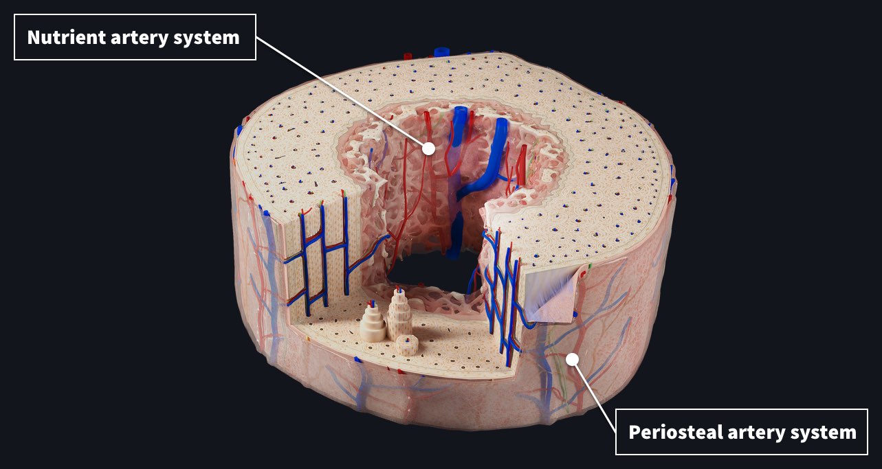
Blood Supply To The Bone Complete Anatomy from cdn.3d4medical.com The wider section at each end of the bone is called the epiphysis (plural = epiphyses), which is filled with spongy bone. Would it be a good thing to show the epiphyseal plate? Compact bone, makes up the dense material in a long section of a bone. Compact bone is the denser, stronger of the two types of bone tissue ( (figure) ). Trust pharmacy 221 massachusetts ave, boston, ma 02115. Bone test anatomy and physiology 12 photos of the bone test anatomy and physiology anatomy and physiology bone lab test, anatomy and physiology bone markings test, anatomy and physiology bone practical test, anatomy and physiology bone tissue test, anatomy and physiology test on bone tissue, bone, anatomy and. The wider section at each end of the bone is called the epiphysis (plural = epiphyses), which is filled internally with spongy bone, another type of osseous tissue. Browse 4,294 bone cross section stock photos and images available, or search for human bone cross section to find more great stock photos and pictures.
This article lists a series of labeled imaging anatomy cases by system and modality.
Why is the marrow red? Looking at a bone in cross section. This photo shows a cross section through bone. The wider section at each end of the bone is called the epiphysis (plural = epiphyses), which is filled internally with spongy bone, another type of osseous tissue. Bone is found in the shafts of long bone and consists of various cylindrical units named as haversian system 47. To the left is muscle tissue, and to the right is bone marrow. Each epiphysis meets the diaphysis at the metaphysis. The wider section at each end of the bone is called the epiphysis (plural = epiphyses), which is filled with spongy bone. In the center of each osteon is the central canal, a space that houses blood vessels and nerves that supply bone. Wing bones were sampled from the right side of skeletally table 1. The outlined area is a cross section of an osteon of compact bone. The information provided on our website, including descriptions of goods, is for reference only. Bone in arm pictures 12 photos of the bone in arm pictures bone cancer arm pictures, pictures of bone cancer in arm, bone, bone cancer arm pictures, pictures of bone cancer in arm
In three dimensions an osteon is cylindrical in shape bone cross section. And why does the marrow stop where it does, and so sharply?